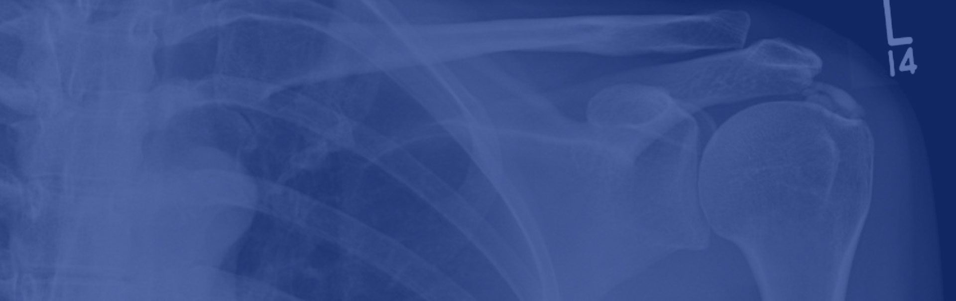
Technique
X-rays are often the first-line investigation in musculoskeletal disorders. The use of X-rays exposes the patient to low dose radiation to produce a 2D image of the body part. It is a regular practice to obtain two separate X-rays often at right angles to each other. X-rays must legally be requested by your consultant, GP or other approved practitioner.


Applications
X-rays show exquisite bony detail and are extremely useful for detecting bone lesions, fractures, arthritis and problems with joint replacements. Soft-tissue features may be poorly defined, so other follow up imaging techniques may be considered to demonstrate any soft-tissue abnormality.
Patient information
No preparation is usually required. If you are between 12 and 55 years old, you may be asked if you could be pregnant as X-rays use small amounts of ionising radiation. X-rays are quick and should not take more than 10 minutes. X-ray images are stored digitally and displayed on a computer screen, enabling our experts to carefully analyse the images and send a report on the findings to the referring doctor.
