Technique
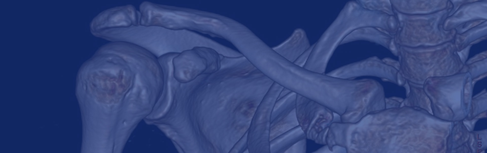
A computerised tomography (CT) scan is a non-invasive scan that uses special circular X-ray equipment that revolves around the body obtaining multiple images of bones and joints from different angles as the patient lies on a moving table. The images are then built into detailed 3 dimensional cross-sectional images by the specialised computer software.


Applications
CT demonstrates fine bony detail and may be used to diagnose and assess fractures and other bone lesions, joints, the spine and to guide some interventional procedures.
It is particularly useful for pre-surgical planning to help our orthopaedic surgical colleagues customise joint replacements. CT may be useful if there are contraindications to MRI examination or if there has been previous surgery with metalwork in the region to be scanned.
Patient information
The whole examination is likely to take about 15 minutes, although the scan itself lasts only a few seconds. CT uses a higher radiation dose than X-rays, but with modern equipment this is kept to the lowest level. Staff must be informed of pregnancy prior to the scan as the procedure uses radiation.
The CT images once obtained in their digital forms are carefully analysed, reconstructed and reported by our experts.
